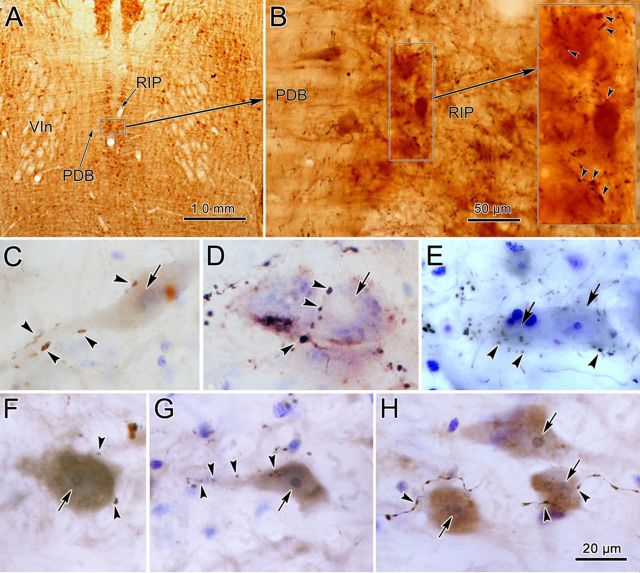
Figure 4.
BDA-labeled and PhaL-labeled cMRF terminals within nucleus RIP. A, Location of RIP in a frontal section counterstained for cytochrome oxidase. Note its position between the exiting abducens nerves (VIn) and flanked by the predorsal bundle (PDB). B, At higher magnification, the arborization of BDA-labeled axons among the RIP cells is demonstrated (arrowheads in inset indicate boutons). C–E, BDA-labeled terminal boutons are closely associated (arrowheads) with Nissl-stained RIP neurons (arrows). F–H, PhaL-labeled cMRF terminal boutons are closely associated (arrowheads) with RIP neurons (arrows). The brown color of the cytoplasm of these cells is due to background staining. Scale bar: H (for C–H), 20 μm.

