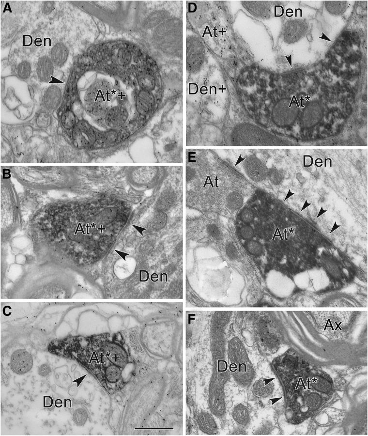
Figure 8.
A–F, Electron micrographs of GABA-positive (A–C) and GABA-negative (D–F) reticuloraphe axons. A–C, Postembedding GABA immunohistochemistry of BDA-labeled cMRF terminals in RIP. Most BDA-labeled axon terminals in RIP were GABA positive (At*+). The BDA-labeled, GABA-positive terminals made symmetric contacts (arrowheads) with GABA-negative dendrites (Den). D–F, Other terminals labeled following cMRF injection were GABA negative (At*). These BDA-labeled profiles made asymmetric contacts (arrowheads) with GABA-negative profiles (Den). At, Unlabeled terminal; Ax, unlabeled axon. Scale bar, 0.5 μm.

