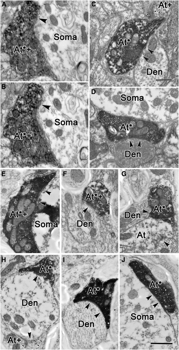
Figure 9.

GABA staining of labeled terminals in the RIP. A, B, A double-labeled axonal terminal (At*+), opposing an unlabeled somatic profile (Soma) was found in two semiserial sections. Interval between adjacent pictures is ∼100 nm. C, D, Electron micrographs of anterogradely labeled, GABA-negative terminals in RIP following a BDA injection in the superior colliculus. Synapses between BDA-positive, GABA-negative terminals (At*) and GABA-negative dendrites (Den) are indicated by arrowheads. In C, a GABA-positive, BDA-negative terminal (At+) is also shown. These profiles contain round, clear vesicles and make asymmetric contacts, although in D no synapse is apparent. Instead, a GABA-negative terminal lies adjacent to a GABA-negative somata (Soma). E–J, PhaL-labeled cMRF terminals in the RIP can be both GABA positive (At*+; E–G) or GABA negative (At*; H–J). GABA-positive terminals that were not labeled by the PhaL injection of the cMRF were also present (At+; H). Small arrows indicate immunogold particles. Scale bars: C (for A–D), 0.5 μm; J (for E–J), 0.5 μm.
