
Figure 9.
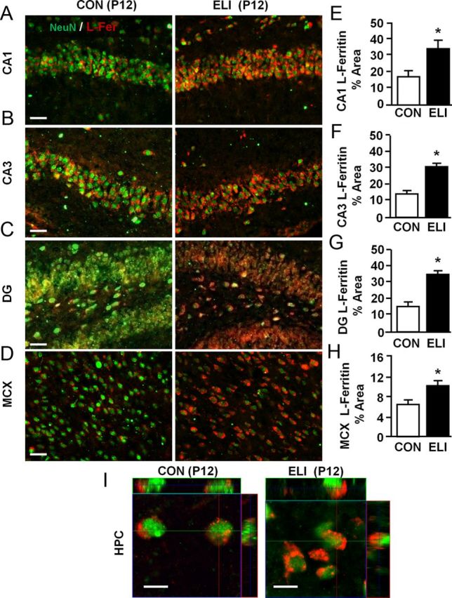
Neonatal infection leads to iron sequestration in neurons before peak myelination. Neonatal mice (P3) were injected subcutaneously with either saline (CON) or E. coli (ELI), and brain tissue from male and female mice was collected at P12 and labeled for NeuN and L-Fer. Representative images of NeuN+ and L-ferritin+ are shown in the (A) CA1, (B) CA3, and (C) dentate gyrus (DG) regions of the HPC, and (D) the CX. Proportional area analyses of L-ferritin labeling in the (E) CA1, (F) CA3, (G) dentate gyrus, and (H) CX are shown. I, Orthogonal projections of interneurons from the CA1 region of the hippocampus for control and ELI mice are provided. Bars represent the mean ± SEM (n = 4). Means with asterisk are significantly different from controls (p < 0.05). Scale bars: A–D, 100 μm; I, 5 μm.