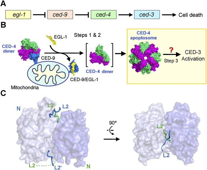
Figure 1.
CED-4-mediated CED-3 activation is a central step to initiate PCD in C. elegans. (A) Four genes—egl-1, ced-9, ced-4, and ced-3—act in a linear fashion to control the onset of PCD in C. elegans. (B) Current model of the linear PCD pathway. In normal cells, CED-4 is sequestered by CED-9, unable to activate the cell-killing caspase CED-3. Upon cell death stimuli, EGL-1 is activated and binds to CED-9. Binding by EGL-1 causes marked conformational changes in CED-9, resulting in the release of CED-4. The released CED-4 dimer oligomerizes to form the CED-4 apoptosome, which in turn facilitates the activation of CED-3. How CED-4 binds CED-3 remains unclear, hindering a mechanistic understanding of CED-3 activation. (C) Structure of the two-chain CED-3 (residues 198–374, 389–503, and C358S). Despite being a monomer in solution, CED-3 was crystallized as an asymmetric dimer, likely due to the high concentrations required for crystallization.

