Figure 4.

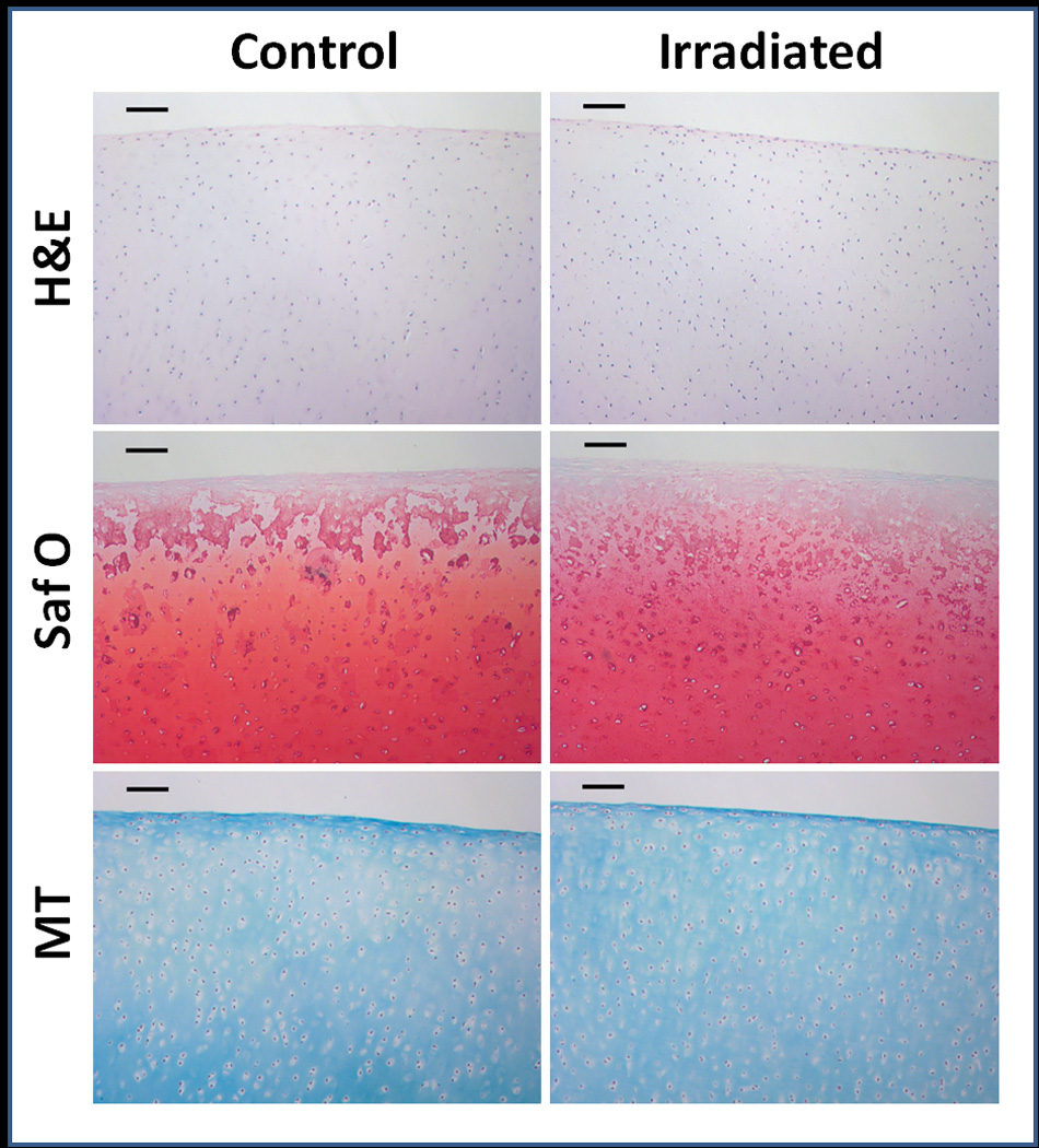
Histological cross sections of articular cartilage explants stained with H&E, Safranin O, and Masson’s trichrome. Qualitatively, there were no differences seen between control and irradiated samples. Scale bars represent 100µm.

Histological cross sections of articular cartilage explants stained with H&E, Safranin O, and Masson’s trichrome. Qualitatively, there were no differences seen between control and irradiated samples. Scale bars represent 100µm.