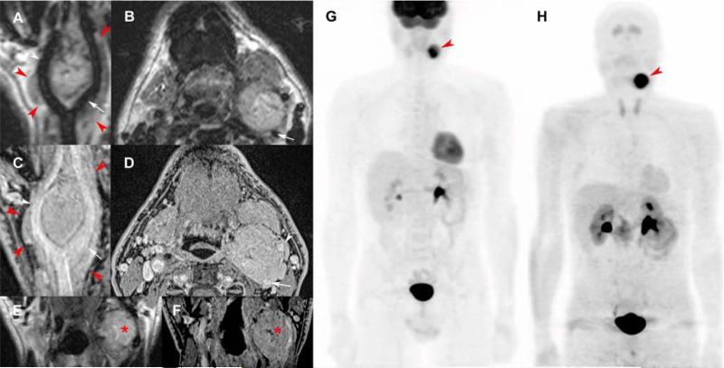
Figure 1. Typical carotid body paraganglioma.
T2-weighted 3D imaging with different flip angle evolutions in curve (A), axial (B), and coronal reconstructions (E); 3D volumetric interpolated fat-saturated (FATSAT) T1-weighted (VIBE) in curve (C), axial (D), and coronal reconstructions (F); 18F-FDG PET/CT (maximal intensity projection (MIP)) (G) and 18F-FDOPA PET/CT (MIP) (H).
MRI shows a “lyre sign” related to a 5-centimeter left carotid body PGL (red arrowheads) arising from the carotid bifurcation, splaying the internal carotid artery (ICE) (A-E, white arrows) and external carotid artery (ECA) (A-D, white arrowheads). Note small central necrosis in the tumor (E, F, red asterisk) and the lack of flow voids due to high temporal resolution of 3D sequences, especially for high-field MRI. 3D reconstructions demonstrate 360° carotid invasion along the common carotid, ICA, and ECA. 18F-FDG PET/CT and 18F-FDOPA PET/CT show a single highly-avid carotid body PGL.

