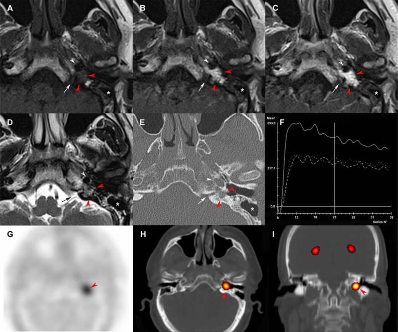
Figure 4. Residual tympanic PGL after surgery.
Axial SE T1-weighted (A), early arterial contrast-enhanced T1-weighted (B), early venous contrast-enhanced T1-weighted (C) and T2- weighted (D), CT scan with a bone algorithm (E), time-intensity curve on dynamic contrast-enhanced MRI (F), axial 18F-FDOPA PET (G), axial 18F-FDOPA PET/CT fusion image (H), coronal 18F-FDOPA PET/CT fusion image (I).
Previous canal wall up tympanomastoidectomy (A-E, asterisk). Residual tympanic PGL (A-E, red arrowheads) located in the petrous apex, posteriorly to the carotid artery (white arrowheads) and lateral to the cochlear aqueduct (arrows). The time-intensity curve shows arterial enhancement of the PGL and enables precise delineation of the lesion prior to sigmoid sinus and nasopharyngeal mucosa enhancements. 18F-FDOPA PET was positive (red arrowheads).

