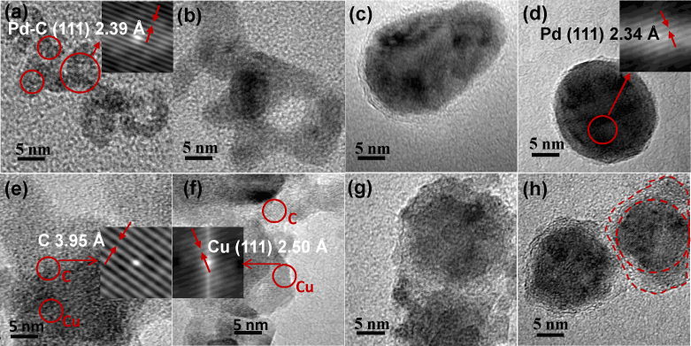
Figure 3. Morphological changes on in-flight sintering.
TEM images showing the compaction and conversion of (a–d) Pd-C and (e–h) Cu-C agglomerates to Pd/MLG and Cu/MLG core shell nanoparticles on sintering. HRTEM image of Pd-C agglomerates (a) showing Pd-C phase which gets converted to Pd phase (d) in the nanoparticle core on sintering at 700°C followed by subsequent cooling. MLG shell around the well formed metal nanoparticles is clearly visible in Figs. c and d. HRTEM images (e–f) of Cu-C nanoparticles show the presence of both Cu and C phases as confirmed by lattice fringes of selected regions in the micrographs (inset of e and f). Graphene shell layer around the well formed metal nanoparticles is clearly visible in Fig. g and h.

