
Figure 1.
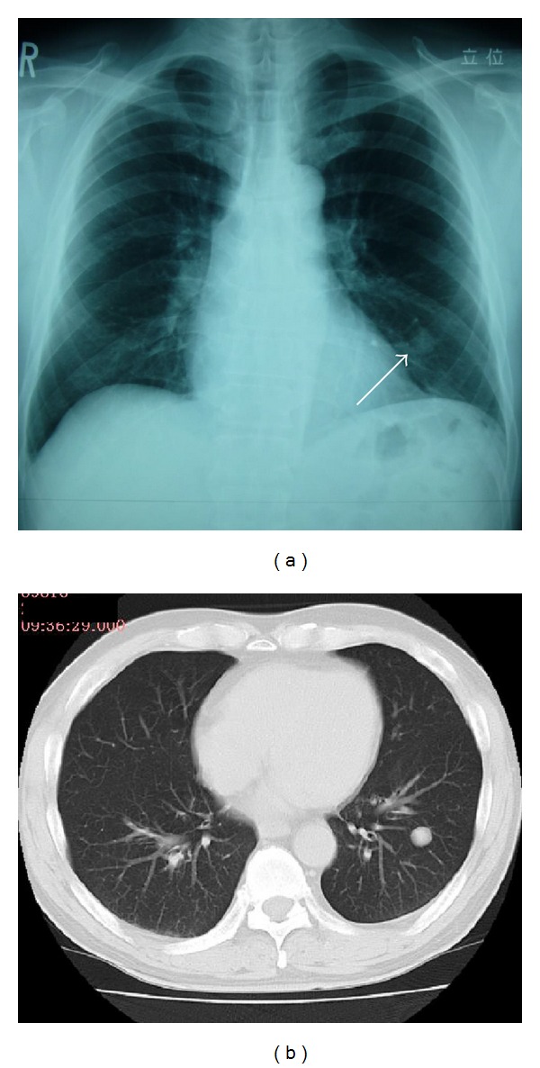
Chest X-ray and CT findings on admission. (a) Simple chest X-ray indicated a smooth, well-circumscribed nodule in the left lower lung field (white arrow). (b) Simple chest computed tomography revealed a nodule measuring 17 mm in diameter and having a well-defined border in S9 of the left lung.