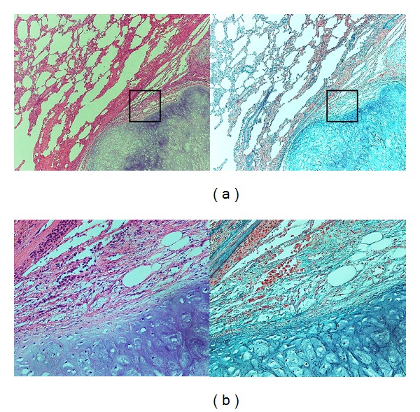
Figure 4.

(a) Histology of the pulmonary hamartoma. The section showing the typical appearance of the pulmonary hamartoma. A lobular mass of mature hyaline cartilage is intimately admixed with fibromyxoid stroma. (Left: Hematoxylin-Eosin stain; ×40. Right: Elastica-Masson stain; ×40). (b) Higher magnification of black box in (a) (Left: Hematoxylin-Eosin stain; ×200. Right: Elastica-Masson stain; ×200).
