Figure 6.
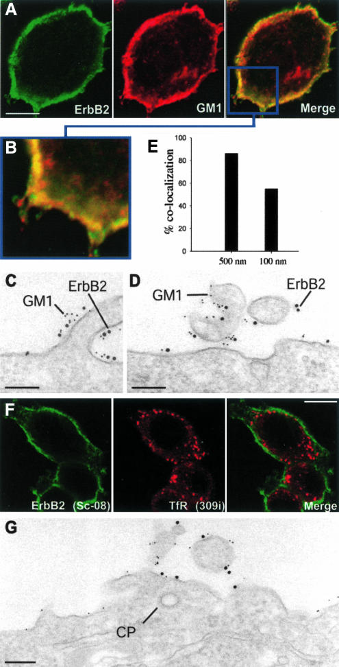
Cholesterol depletion does not lead to internalization of ErbB2. (A) Confocal images of an SKBR3 cell stained for ErbB2 and GM1. The merged image indicates a high degree of colocalization at the cell surface. (B) Higher magnification of a part of the merged image. (C and D) EM pictures of cells double labeled for ErbB2 (10-nm gold) and GM1 (5-nm gold). (E) Quantification of the gold labeling showing that most ErbB2 gold particles have neighboring GM1 particles both within the 500- and the 100-nm range. (F) Confocal images of cholesterol-depleted cells (mβCD) double labeled for ErbB2 and TfR. No internalized ErbB2 is observed. (G) EM immunogold double labeling for ErbB2 (10 nm) and GM1 (5 nm) of a cholesterol-depleted cell showing that ErbB2 have not been removed from the protrusions or entered clathrin-coated pits (CP). Bar, 10 μm (A), 200 nm (C and D), 10 μm (F), and 200 nm (G).

