Figure 1.
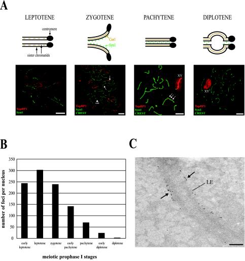
Localization of TopBP1 protein during meiotic prophase I in mouse testis. (A) The four stages of meiotic prophase I (top). Fluorescence microscopy of mouse meiotic chromosomes stained with rabbit anti-TopBP1 antibody (red), mouse anti-Syn1 and human CREST anticentromere serum (green; bottom). In leptotene, TopBP1 is associated with newly formed chromosome cores. In zygotene, TopBP1 is mainly located to chromosome cores that are not completely synapsed (arrows). The arrowhead indicates two single centromeres (cen). In the fully synapsed pachytene stage there are fewer TopBP1 foci on autosomes but the X-Y pair is brightly stained (XY). The green and red images are slightly offset to show the association of TopBP1 foci with the chromosome cores (arrows). In diplotene, TopBP1 is staining only the X-Y pair (XY). Bars, 20 μm. (B) Average number of TopBP1-containing foci during the different stages of the meiotic prophase I. Thirty-two nuclei in total (at least 3 nuclei per stage) stained with rabbit anti-TopBP1 antibody were analyzed. (C) Electron micrograph of a full pachytene SC with many 10-nm gold grains marking TopBP1 sites (arrows) at the lateral elements. LE, lateral element. Magnification, 20.000×. Bar, 200 nm.

