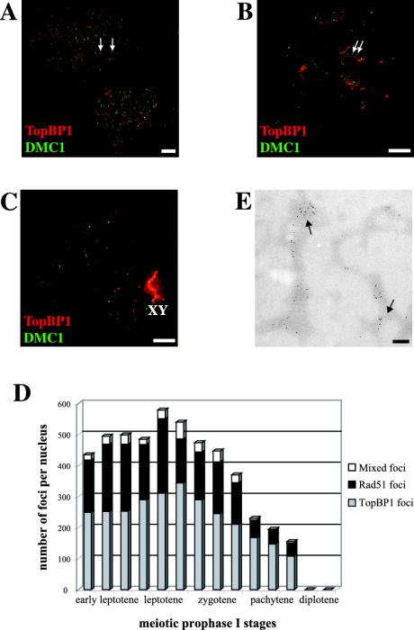
Figure 2.
Coimmunostaining of TopBP1 and Dmc1 proteins in different stages of mouse meiotic prophase I. A, B, and C are stained with rabbit anti-TopBP1 (red) and mouse anti-Dmc1 (green). (A) Two leptotene nuclei showing numerous bright TopBP1 and Dmc1 foci, ∼10% of them colocalizing (yellow foci, arrows). (B) One zygotene nucleus showing bright TopBP1 staining on yet unsynapsed chromosome cores. The number of Dmc1 foci is lower than in leptotene, but there is a higher degree of colocalization (20%) of the two types of foci (arrows). The red and green images are slightly offset to better show this association. (C) One pachytene nucleus showing very bright TopBP1 staining on the X-Y pair (XY) and a few foci on the autosomes. Dmc1 protein is coming off the chromosome cores at this stage, as shown by the weak green signal. Bars, 20 μm. (D) Numbers of Rad51/Dmc1, TopBP1, and mixed foci per nucleus that are associated with chromosome cores/SCs at successive developmental stages, based on 14 completely analyzed nuclei. A small proportion of the foci contain all three proteins represented by the white segments of the bars. (E) Immunoelectron micrograph of chromosome cores stained with 5- and 10-nm gold particles marking Dmc1 and TopBP1 sites, respectively. Bar, 200 nm.

