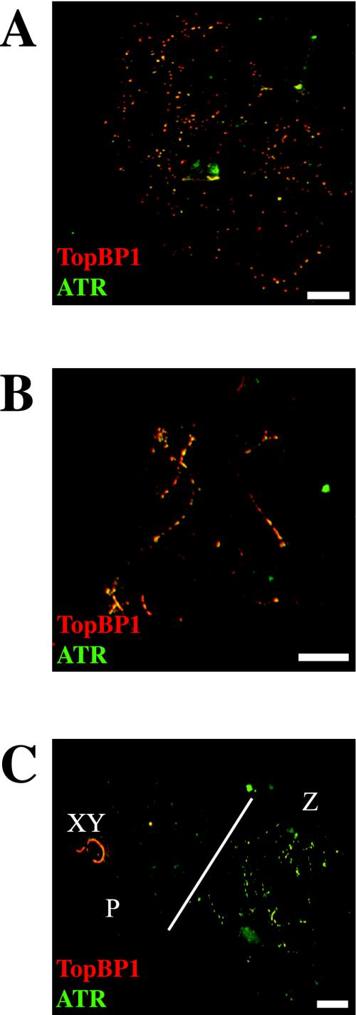
Figure 3.
Coimmunostaining of TopBP1 and ATR proteins in different stages of mouse meiotic prophase I. A, B, and C are stained with rabbit anti-TopBP1 (red) and sheep anti-ATR (green) antibodies. (A) One leptotene nucleus showing many yellow bright foci, demonstrating that TopBP1 and ATR colocalize in most, if not all, foci. (B) One zygotene nucleus showing an intense yellow staining of yet unsynapsed regions. (C) Two different nuclei are shown. The one on the right (Z) is a zygotene nucleus showing the same pattern as in B. The one on the left (P) is a pachytene nucleus showing TopBP1 on the X-Y pair (XY) and a few weak foci on the autosomes. In this nucleus, ATR is only present on the X-Y pair. Bars, 20 μm.

