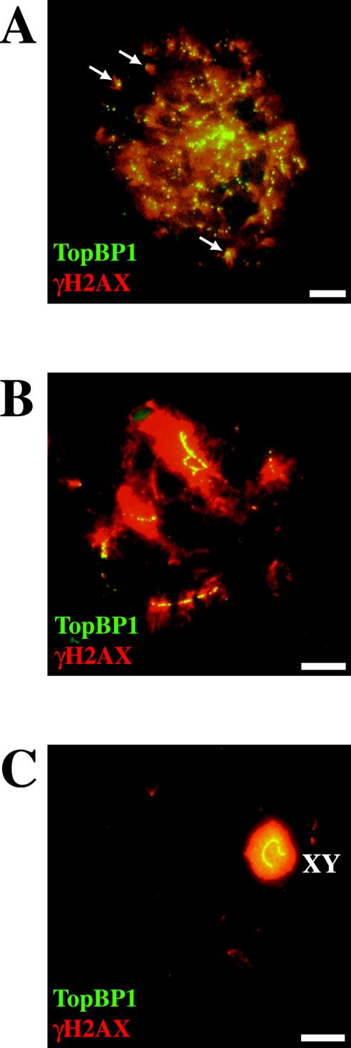
Figure 4.
Coimmunostaining of TopBP1 and γ-H2AX proteins in different stages of mouse meiotic prophase I. A, B, and C are stained with rabbit anti-TopBP1 (green) and mouse anti-γ-H2AX (red). (A) One late leptotene nucleus showing the colocalization of the two proteins (yellow). All TopBP1 foci colocalize with γ-H2AX, but there are many γ-H2AX–positive domains that do not contain TopBP1 foci. Arrows indicate isolated γ-H2AX domains containing TopBP1 foci. (B) One zygotene nucleus showing TopBP1 staining on yet unsynapsed regions that are also positive for γ-H2AX staining. (C) A late pachytene nucleus showing TopBP1 and γ-H2AX uniformly coating the sex chromosomes. Bars, 20 μm.

