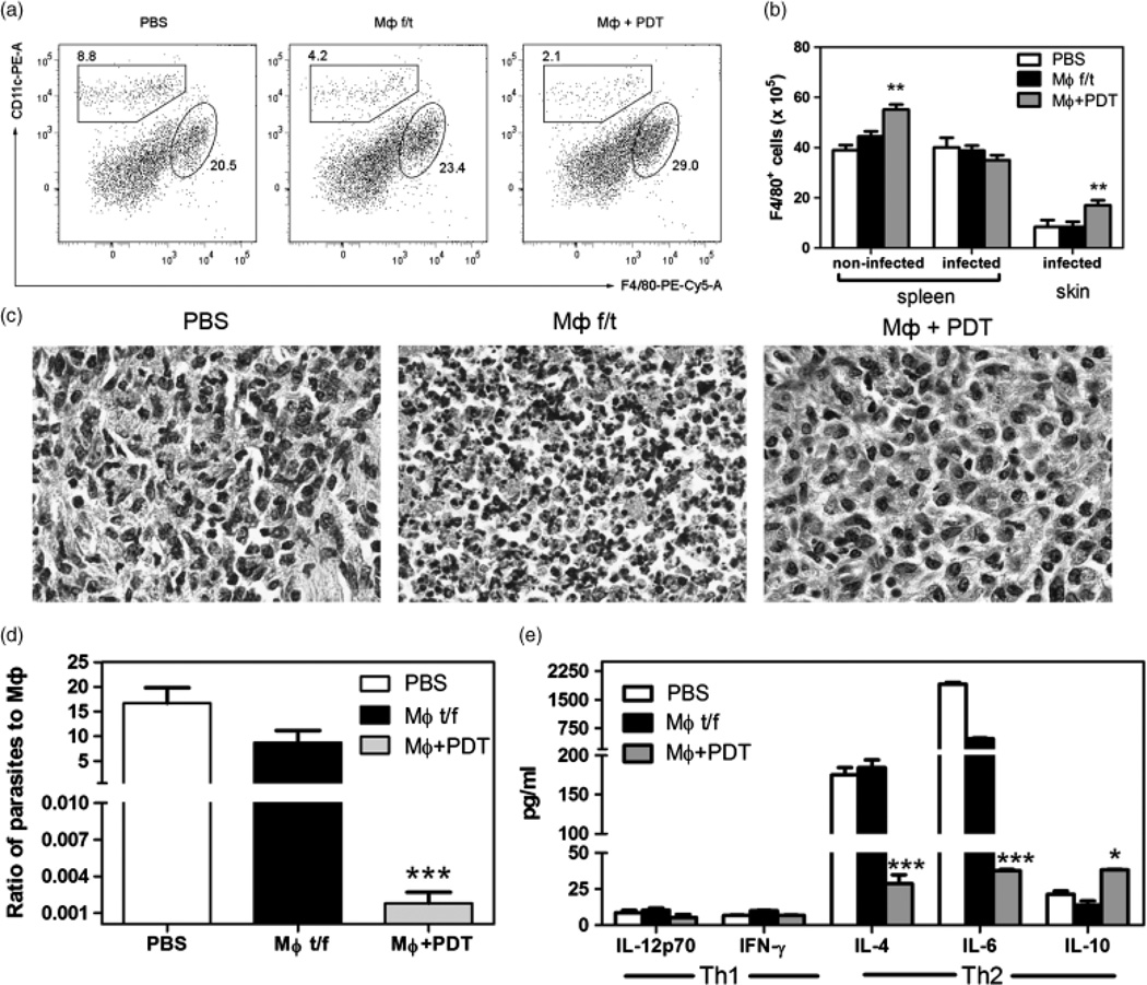
Fig. 3.
Vaccination with PDT-treated, apoptotic MΦ increased the number of MΦ and suppressed Th2 immune response. (a) Dot plots for F4/80 and CD11c expression in the spleens of vaccinated non-infected mice. (b) Absolute numbers of F4/804cells in and ears with the CL lesions in vaccinated non-infected or infected mice. Mean ± SD for each group. ** P < 0.01 in comparison with MΦ f/t. (c) Histological picture of the CL lesions of vaccinated mice 3 weeks after infection. H&E, × 800. (d) Ratio of parasites to macrophages in the CL ears of vaccinated mice 3 weeks after infection. (e) Cytokine production in the CL lesions of vaccinated mice 3 weeks after infection. **P < 0.05, ** P < 0.001 in comparison with PBS.

