Fig. 4.
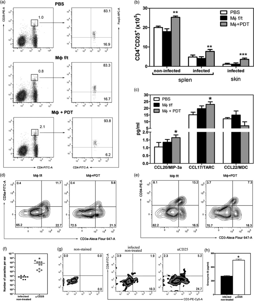
Vaccination with PDT-treated, apoptotic MΦ affected the number of Treg, CD8 Tand NKT cells (vaccinated mice 3 weeks after infection). (a) Percentage of CD4+ CD25high in the spleens. (b) The absolute number of CD4+ CD25+ in the spleen and skin. **P < 0.01, ***P < 0.001. Five mice per groups of three independent experiments. (c) Chemokines in the CL lesions. *P < 0.05. (d) CD3 and CD8 in the spleens of vaccinated non-infected mice. Five mice per group. (e) CD3 and CD94 in the spleens of vaccinated non-infected mice. CD25-depletion in infected mice. (f) Parasite load. *P < 0.001 (g) CD3 and CD8a in the CL lesions after CD25 depletion. (h) The granzyme B in the CL lesions after CD25 depletion. Ten ears per sample. *P < 0.05.

