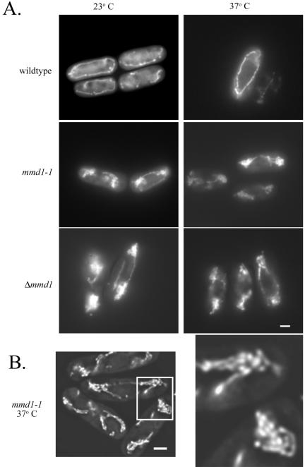
Figure 1.
Mutant mmd1 cells display abnormal mitochondrial morphology and distribution. Wild-type (MYP115), mmd1-1 (MYP106), and Δmmd1 (MYP105) were grown in YES medium at 23°C and incubated for 4 h at 23°C or 37°C. (A) Mitochondrially targeted GFP was visualized in wild-type, mmd1-1 and Δmmd1 cells by using fluorescence microscopy. (B) Mitochondrially-targeted GFP was visualized in mmd1-1 cells incubated for 4 h at 37°C with fluorescence microscopy on an Olympus IX-70 inverted microscope. Images were taken and deconvolution was performed on 64 optical sections with a step of 0.2 μm by using Delta Vision 2.10 software (Applied Precision, Issaquah, WA). Bar, 2 μm.

