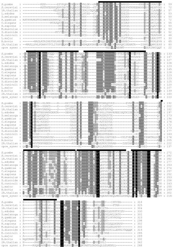
Figure 5.
Alignment of S. pombe Mmd1p and its homologues in diverse organisms. Amino acid sequences were aligned using the ClustalX program (Thompson et al., 1997) and the alignment saved in GCG/MSF format. The alignment file was processed with the GeneDoc program (http://www.psc.edu/biomed/genedoc). Shading levels represent 100% (darkest), >80%, >60%, and <60% conservation of amino acid residues among homologues for a given position. Bars over the Mmd1p sequence of S. pombe show the position of the EZ-HEAT motifs, as identified by the SMART database (Schultz et al., 2000). The residue mutated in the mmd1-1 strain is indicated by the Λ.

