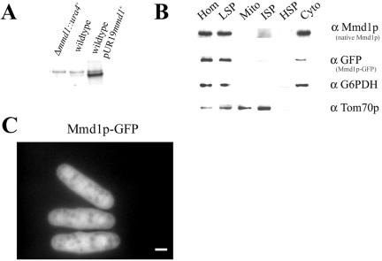
Figure 7.
Mmd1p is a cytosolic protein. (A) Cellular homogenates were prepared from Δmmd1 (MYP105) and wild-type (FY254) cells and from wild-type cells harboring a multicopy plasmid (pUR19mmd1+) encoding mmd1+ (MYP114). Proteins were analyzed by SDS-PAGE on an 8–16% gradient gel and immunoblotted with antibodies against Mmd1p. Each lane contained 25 μg of total protein. (B) Subcellular fractions were prepared from cells harboring a single copy of the gene encoding Mmd1p-GFP (MYP112). Proteins were separated by SDS-PAGE and analyzed by immunoblotting. Fractions were probed with anti-Mmd1p, anti-GFP, anti-Glucose-6-phosphate dehydrogenase (G6PDH), a cytosolic marker, and anti-Tom70p, a mitochondrial marker. Subcellular fractions are: HOM, total cell homogenate; LSP, low-speed pellet; Mito, mitochondrial fraction; ISP, intermediate-speed pellet; HSP, high-speed pellet; and Cyto, cytosolic fraction. Each lane contained 10 μg of total protein. (C) Microscopic localization of Mmd1p. Cells containing a single copy of Mmd1p-GFP (strain MYP112) were grown on YES medium at 30°C and analyzed by fluorescence microscopy. Bar, 2 μm.

