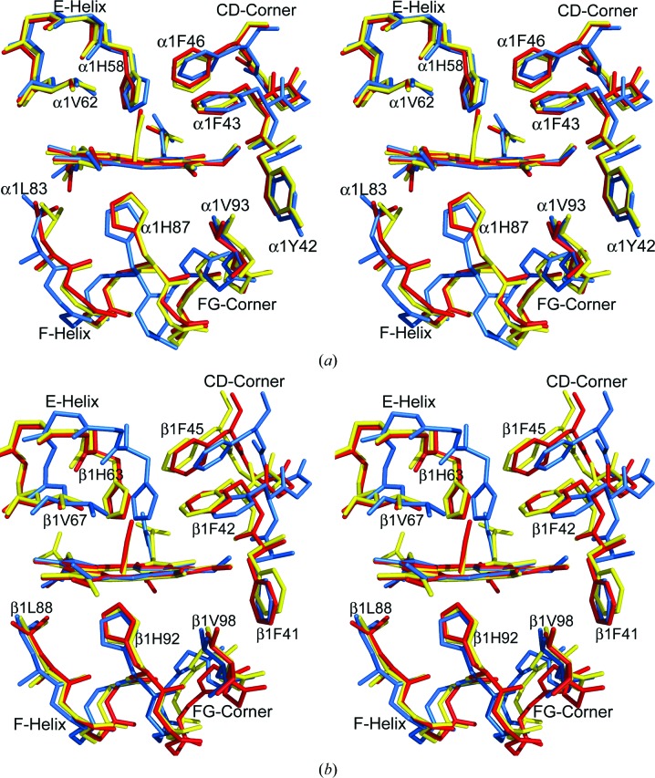
Figure 2.
Stereoview of the heme environment of deoxy T (PDB entries 2hhb or 2dn2; cyan), CO-ligated R (PDB entries 1aj9 or 1ljw; red) and CO-ligated Hb ζ2βs 2 (yellow) structures after superposing the α1 (ζ1) subunit or β1 subunit by least-squares fitting of the porphyrin pyrrole atoms. Note the different positions of the F helix, E helix, EF corner and CD corner. (a) The α1 (ζ1) heme. (b) The β1 heme (βs1).

