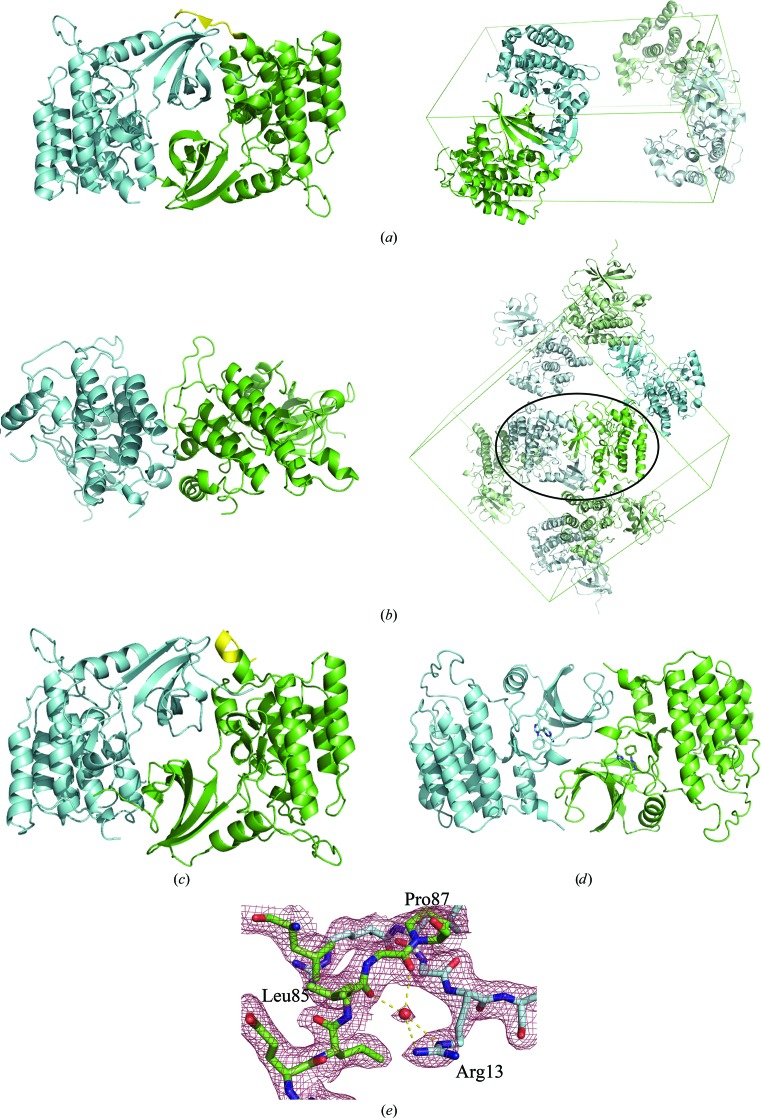
Figure 5.
Asymmetric units and crystal packing in CK1δ structures. (a) Left, the asymmetric unit of P21 CK1δ (1–299). Residues 293–299 of chain A are shown in yellow. Right, the unit cell of P21 CK1δ (1–299). The symmetry-related asymmetric unit is shown in pale green/cyan. (b) Left, the asymmetric unit of PDB entry 1cki. Right, the unit cell of PDB entry 1cki. Symmetry-related asymmetric units are shown in pale green/cyan. The circled crystal-packing interface corresponds to the dimer interface observed in P21 CK1δ (1–299). (c) Closer view of the two 1cki monomers related by the circled crystal contact. The C-terminus of chain A (yellow) adopts a different conformation than in the asymmetric unit of P21 CK1δ (1–299). (d) The asymmetric unit of PDB entry 3uzp, which despite crystallizing in the same space group has a completely different dimer interface to P21 CK1δ (1–299) owing to the presence of the inhibitor PF670462 in the ATP-binding cleft (shown as a stick model), which blocks some of the interactions between monomers observed in the P21 CK1δ (1–299) asymmetric unit. (e) Close-up of the dimer interface in P21 CK1δ (1–299), showing the interaction between the side chain of Arg13 and the hinge region of the other molecule. Unweighted 2F o − F c electron density is shown contoured at 1.0σ.

