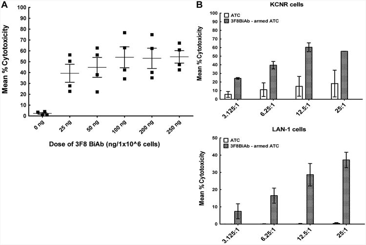
Fig. 4.
A: Arming dose titration of 3F8BiAb. Cytotoxicity mediated by ATC armed with the 3F8BiAb at doses of 25, 50, 100, 200, and 250 ng per 1 × 10⁶ cells was measured in 51Cr-release assay against KCNR neuroblastoma cells. Controls included unarmed ATC (0 ng). E:T of 25:1 was used for these experiments. Mean % cytotoxicity ± SEM for four healthy donors (■) is shown for each dose of the BiAb. *P < 0.01 and **P < 0.007 as analyzed by paired t-test. B: Cytotoxicity of 3F8BiAb armed ATC increases with E:T ratio. ATC generated from healthy donors (n = 2) were armed with 100 ng/1 × 106 cells of 3F8BiAb (
 ) and co-cultured at four different E:T ratio; overnight at 37°C in 96-well flat-bottom plates containing 51Cr-labeled KCNR (upper panel) or LAN-1 (lower panel) target cells. Unarmed ATC were used as controls (□). Mean % cytotoxicity was calculated and error bars represent SD from two experiments.

