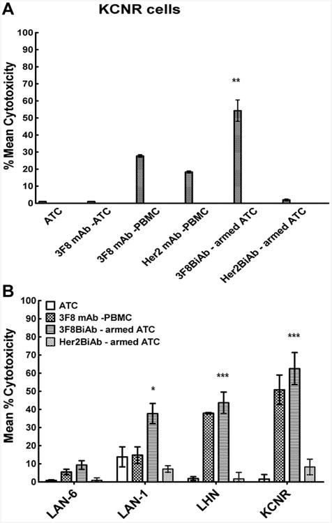
Fig. 5.
A: 3F8 mAb dependent ADCC against neuroblastoma cells. 51Cr labeled target cells were co-cultured in the presence of unarmed ATC, 3F8 mAb plus ATC, 3F8 mAb plus PBMC and armed ATC with 3F8 BiAb, controls comprised of Her2 mAb in the presence of PBMC and Her2BiAb-armed ATC. A standard 18-hour chromium release cytotoxicity assay was performed as described in materials and methods. E:T of 25:1 was used. Representative donor for neuroblastoma (KCNR) cells is shown. Error bars represent SD of two replicates for each condition. **P < 0.01 for KCNR cells versus 3F8mAb plus PBMC control. B: 3F8BiAb-armed ATC kill GD2-expressing neuroblastoma cells in co-culture. LAN-6, LAN-1, LHN, and KCNR neuroblastoma cell lines were exposed to ATC armed with 100 ng/1 × 106 cells of 3F8BiAb (
 ) in a standard 51Cr release assay. ATC alone (□), 100 ng/well of 3F8 mAb combined with PBMC (
 ) and Her2Bi-armed ATC (
 ) were used as controls. E:T was 25:1. Mean % cytotoxicity ± SEM of six donors with each condition done in duplicates is shown. *P < 0.05 and ***P < 0.001 versus the unarmed ATC control.

