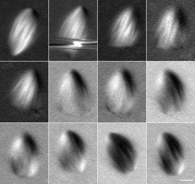
Figure 5.
Large spindle fragments are able to reform complete spindles after cutting. Spindles were cut as above and imaged by polarization microscopy for 10-20 min. Frames from a time-lapse image are shown; the interval between frames was 80 s. By ∼10 min, a recognizable spindle pole has formed where the cut edge was originally produced. This spindle rotated counterclockwise during the second part of the time-lapse image. The smaller spindle fragment was brushed away by the lower cutting needle. Bar, 10 μm.

