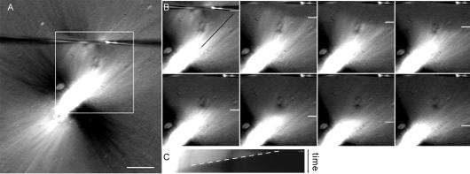
Figure 6.
Astral microtubules depolymerize rapidly after cutting. Astral microtubules were induced to polymerize by the addition of α-MCAK antibody, in order to allow their visualization at 20× magnification. (A) Spindle treated with α-MCAK antibody, imaged by polarization microscopy. Contrast was maximized to emphasize astral microtubules. While microtubules oriented at 45° angles relative to the polarizer are most evident (white and black), the spindle is encircled by astral microtubules at all angles. The cutting needles are visible near the top of the frame. Bar, 20 μm. (B) Frames from a time-lapse image showing the boxed region of the spindle in A. The position of the depolymerization front is shown by the white line, and the line used to generate the kymograph is shown by the black line along the fibers. See Supplemental Movie 6. (C) Kymograph of the region in B, showing the movement of the depolymerization front over time (dashed line, position vs. time).

