
Figure 3.
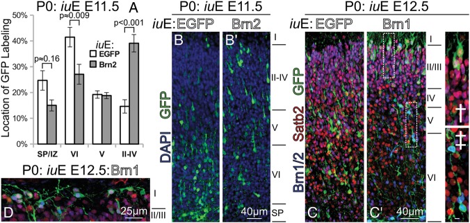
Brn1/2 overexpression confers cell-autonomous pial-directed neural migration to superficial positions. (A) Quantification of the position of GFP labeling at P0 in electroporations of Brn2 versus EGFP performed at E11.5. (B and B′) As shown in (A), Brn2-electroporated cells had a strong preference for superficial laminar positions (B′) compared with EGFP-only electroporated cells (B); this came at the expense of layer VI and subplate positioning. (C and C′) Similar electroporations, although performed at E12.5 with Brn1, result in a gross change in laminar position at P0. Most GFP+(green) cells in EGFP electroporations at E12.5 (C) assume positions predominantly within layer V and VI. Forced expression of Brn2 at E12.5 (C′) results in the layer II/III-targeted migration of GFP+(green)/Satb2+(red) cells at P0, many reaching positions just beneath layer I(C′†). Dashed boxes in (C′) indicate regions of interest enlarged in † and ‡. Some of the highest Brn1-expressing cells remained in the deep layers (C′‡); oddly, these cells labeled very strongly with Brn2 antibody (blue), and were Satb2-. Scale bar in (C′‡), 5 μm. (D) In the caudal cortex, Brn1/2-mediated superficially targeted migration was so robust that many Brn1-electroporated GFP+ (green) electroporated cells were seen in layer I at P0; those cells were immunolabeled, and were positive for Brn2 (blue) and Satb2 (red).