Figure 1.
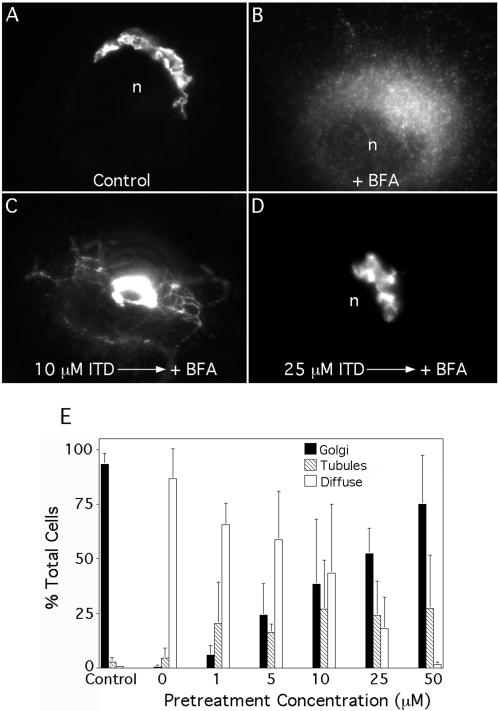
ITD inhibits BFA-stimulated formation of membrane tubules from the Golgi complex and retrograde trafficking to the ER in a dose-dependent manner. Clone 9 cells were washed three times with MEM (without serum), pretreated with various concentrations of ITD for 10 min, and then incubated with BFA (5 μg/ml) in the continuous presence of ITD (as appropriate) at 37°C. Cells were then fixed and stained for immunofluorescence of ManII. (A-D) Immunofluorescence micrographs: (A) Untreated cells with a typical juxtanuclear Golgi complex; (B) BFA treatment induced the redistribution of ManII to a diffuse, ER pattern and the complete loss of the central Golgi complex; (C) cell pretreated with 10 μM ITD shows an intermediate effect with membrane tubules and some central Golgi staining; (D) cell pretreated with 25 μM ITD in which BFA-induced redistribution to the ER was completely inhibited. (E) Quantitation of immunofluorescence results in which cells staining for ManII were scored as having a typical Golgi complex (as in A and D), diffuse ER staining (B), or tubule formation (C). Results show the mean ± 1 SD (n ≥ 3 for all data points). n, nucleus.

