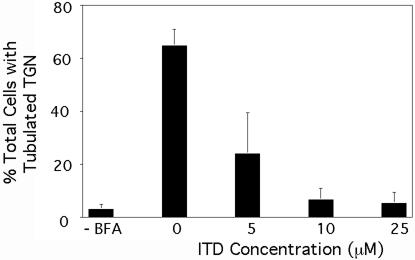
Figure 5.
BFA-induced tubule formation from the TGN is inhibited by ITD. Clone 9 cells were washed in MEM (without serum), pretreated with varying concentrations of ITD, and then subsequently incubated with BFA (5 μg/ml) in the continuous presence or absence of ITD as appropriate for 15 min at 37°C. Cells were then fixed and processed for immunofluorescence localization of the CI-M6PR. Quantitative results show the percentage of cells with extensive TGN tubules. Results show the mean ± 1 SD (n = 3).

