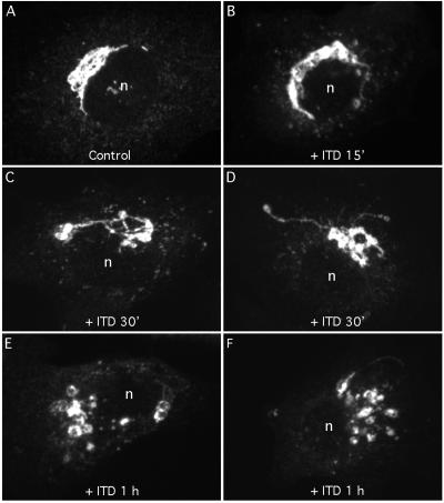
Figure 6.
ITD alone causes fragmentation of the juxtanuclear Golgi ribbon. Clone 9 cells were washed with MEM (minus serum) and then incubated for various periods of time with 25 μM ITD. Cells were then fixed and stained with anti-ManII antibody and visualized by immunofluorescence and confocal microscopy. (A) Untreated cells; (B) cells treated for 15 min; (C and D) cells treated for 30 min; and (E and F) cells treated for 1 h. Each micrograph represents the composite image obtained from ∼20, 0.2-μm confocal slices.

