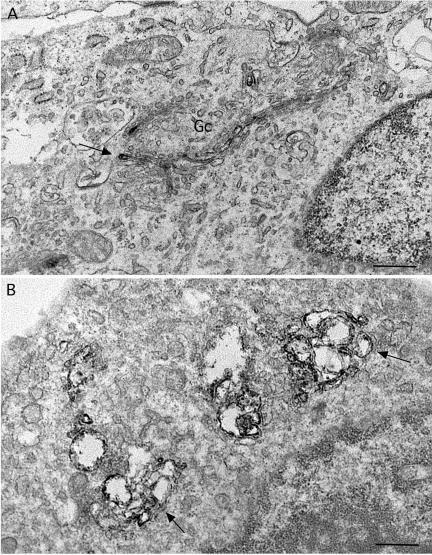
Figure 7.
Immunoperoxidase localization of ManII at the electron microscopic level in ITD-treated cells reveals dilated Golgi cisternae. (A) Control cells with a Golgi complex (Gc) consisting of stacks of flattened cisternae. The electron-dense immunoreaction product of ManII staining is typically found in one or two medial cisternae (arrow). (B) Cells treated with ITD (25 μM) for 1 h show extensively dilated cisternae that contain immunoreaction product (arrows). The dilated cisternae still appear to be stacked together in clumps. Bars, 0.5 μm.

