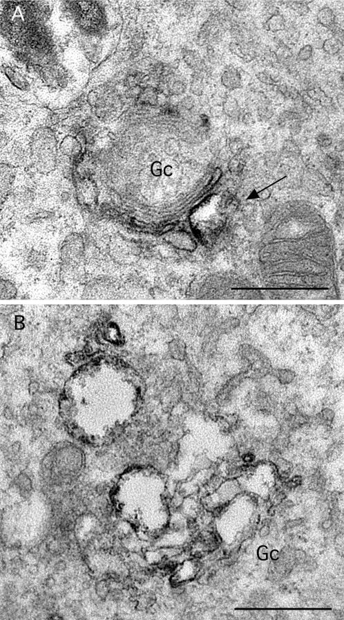
Figure 8.
Higher magnification views of Golgi complexes in ITD-treated cells. (A) In cells treated with ITD (25 μM) for 15 min Golgi complexes (Gc) were often fairly normal but with some dilated cisternae (arrow), in this case, one that contains ManII immunoreaction product. (B) In cells treated for 1 h, all cisternae appeared to be swollen, although they are still found within a stacked unit. Bars, 0.5 μm.

