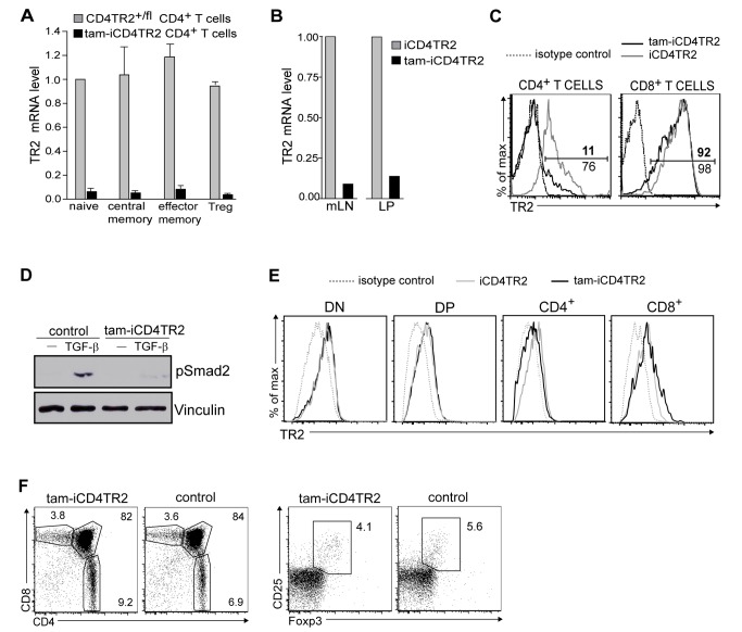
Figure 1. Validation of CD4-CreERt2/tamoxifen-mediated TR2 ablation.
(A) Quantitative RT-PCR of TR2 mRNA in FACS-sorted splenic CD4+ T cell subsets. These data are representative results of three independent experiments. (B) Quantitative RT-PCR of TR2 mRNA in FACS-sorted CD4+ T cells from mesenteric lymph nodes and lamina propria. These data are representative results of three independent experiments. (C) Flow cytometric analysis of TR2 expression by CD4+ and CD8+ T cells from peripheral blood. These data are representative results of four independent experiments. (D) Western blot analysis of pSmad2 and Vinculin in lysates of magnetically purified splenic CD4+ T cells from tam-iCD4TR2 and control mice 2 wk p.a.. The cells were cultured for 40 min in the presence of antiCD3/CD28 either with or without 20 ng/ml TGFβ-1. These are representative data of two independent experiments. (E) Flow cytometric analysis of TR2 expression by thymocytes 2 wk p.a. These data are representative results of two independent experiments. (F) Flow cytometric analysis of expression of CD4 and CD8 by thymocytes (left panel) as well as Foxp3 and CD25 by CD4+ SP thymocytes (right panel) from tam-iCD4TR2 and control mice at 2 wk p.a. These data are representative results of three independent experiments.

