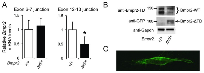
Figure 1. Bmpr2 expression in PaSMCs obtained from WT or Bmpr2 Δtd/+ mice.

(A) Levels of Bmpr2 mRNA were measured in WT (Bmpr2+/+) or Bmpr2 Δtd/+ PaSMCs by qPCR using hydrolysis probes for Bmpr2 exon junctions 6–7 and 12–13. Bmpr2 mRNA levels were normalized to Gapdh and expressed as the fold-change relative to Bmpr2 +/+ PaSMCs. *P < 0.01 compared to Bmpr2 +/+ PaSMCs. (B) Immunoblots prepared from lysates of Bmpr2 +/+ and Bmpr2 Δtd/+ PaSMCs were incubated with an antibody directed against the tail domain of Bmpr2 to detect Bmpr2‑WT or with an anti-GFP antibody to detect Bmpr2‑ΔTD. Immunoblots were subsequently incubated with an antibody directed against Gapdh as a control for protein loading. (C) Confocal microscopy image of a PaSMC transiently transfected with a plasmid directing expression of Bmpr2 Δtd and reacted with an anti-GFP antibody showing localization of Bmpr2‑ΔTD at the cell membrane.
