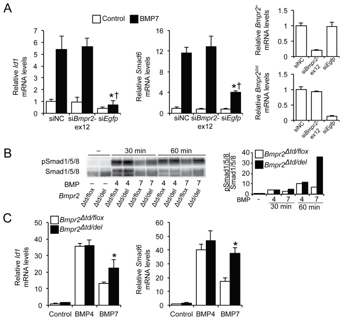
Figure 3. Bmpr2‑ΔTD contributes to BMP7 signaling in Bmpr2 Δtd/+ PaSMCs.
(A) Bmpr2 Δtd/+ PaSMCs were transfected with negative control siRNA (siNC), siBmpr2‑ex12, or siEgfp (30 nM). After 48 h, the ability of BMP7 (10 ng/ml for 1.5 h) to induce Id1 and Smad6 mRNA expression was measured by qPCR, normalized to Gapdh and expressed as fold-change relative to Bmpr2 Δtd/+ PaSMCs transfected with siNC. *P < 0.01 compared to siNC group treated with BMP7, † P<0.01 compared to siBmpr2‑ex12 group treated with BMP7. Efficiency of silencing Bmpr2 + (siBmpr2‑ex12) and Bmpr2 Δtd (siEgfp) transcripts was measured by qPCR. (B) Bmpr2 Δtd/flox and Bmpr2 Δtd/del PaSMCs were treated with BMP4 or BMP7 (10 ng/ml) for 30 and 60 minutes, upon which the activation of Smad1/5/8 was evaluated by immunoblotting. Quantification of the Smad1/5/8 activation is plotted as the ratio of pSmad1/5/8 to total Smad1/5/8. (C) The ability of BMP4 or BMP7 to induce Id1 and Smad6 gene expression in Bmpr2 Δtd/flox and Bmpr2 Δtd/del PaSMCs was measured by qPCR, normalized to Gapdh and expressed as fold-change relative to untreated Bmpr2 Δtd/flox PaSMCs. *P < 0.01 compared to Bmpr2 Δtd/flox PaSMC group treated with BMP7.

