
Figure 2. NMR structure of Bcl-xl (residue 1-217, ∆45-84) in complex with Bak BH3 domain(residue 572-587) from PDB ID ‘1BXL’. Different helices of Bcl-xl (h1-h8) are shown in different colors.
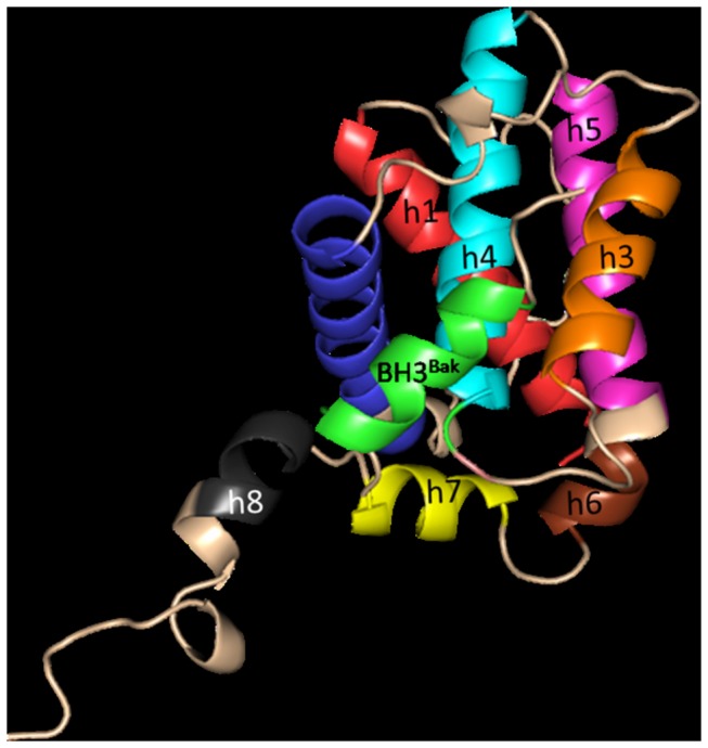
BH3Bak is shown in green.
Official websites use .gov
A
.gov website belongs to an official
government organization in the United States.
Secure .gov websites use HTTPS
A lock (
) or https:// means you've safely
connected to the .gov website. Share sensitive
information only on official, secure websites.
BH3Bak is shown in green.