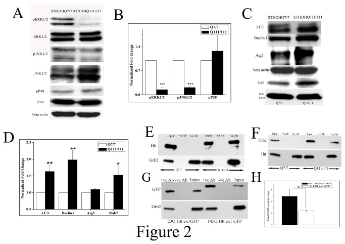
Figure 2. Upregulated Grb2 interacts with mutant Htt and deviates from its normal function.
A. Western blot analysis for the expression of phospho ERK1/2, ERK1/2, phospho JNK1/2, JNK1/2, phospho p38, p38 and beta actin levels in STHdhQ7/7 and STHdhQ111/111 cells. B. Bar diagram of densitometric analysis for three independent (n=3) experiments as shown in panel A the values of phospho ERK1/2 was normalized to that of total ERK1/2 levels, phospho JNK1/2 was normalized to total JNK and phospho p38 level was normalized to that of total p38 level (for pERK1/2 and pJNK1/2 ;p<0.001). C. Western blot analysis for the expression of LC3, beclin1, Atg5, Rab7 and beta actin in STHdhQ7/7 and STHdhQ111/111 cells. D. Bar diagram of densitometric analysis of three (n=3) different experiments as shown in panel C, beta actin taken as internal control (p<0.01 for LC3 and Beclin1, p<0.05 for Rab7). E. Immunoprecipitation experiment (n=3) of Grb2 with Htt in STHdhQ7/7 and STHdhQ111/111 cells. Cell extract was pulled with anti-Grb2 antibody and the pulled down protein was probed with Htt antibody in 6% SDS-PAGE and for the pulled Grb2 was run in 12% SDS-PAGE with same sample and probed with anti Grb2 antibody. F. Immunoprecipitation experiment (n=3) of Grb2 with Htt in STHdhQ7/7 and STHdhQ111/111 cells. Cell extract was pulled with anti-Htt antibody and the pulled down protein was probed with anti Grb2 antibody in 12% SDS-PAGE and for the pulled Htt was run in 6% SDS-PAGE with same sample and probed with anti Htt antibody G. Immunoprecipitation experiment (n=3) with Neuro2A cells transfected with 23Q and 145Q Httex1 GFP respectively and pulled with anti-Grb2 antibody, probed with anti GFP antibody. H. Bar diagram of densitometric analysis of three independent (n=3) as shown in panel G.

