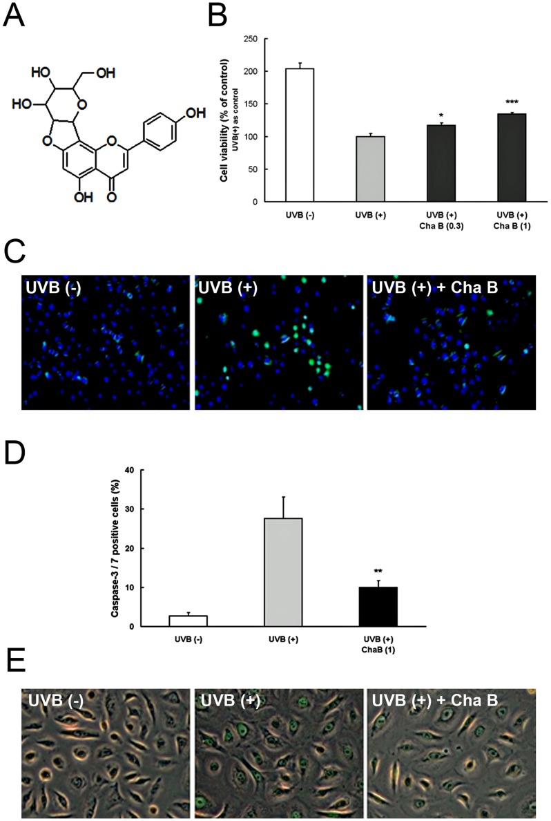
Figure 1. Chafuroside B attenuated DNA damage, cell damage, and apoptosis in UVB-exposed human keratinocytes.
A Chemical structure of chafuroside B. B NHEK were irradiated with UVB (20 mJ/cm2), and then treated with chafuroside B (0.3 and 1 µM). Alamar blue assay was used to evaluate cell viability at 48 h after treatment. C, D NHEK were irradiated with UVB (20 mJ/cm2), and then treated with chafuroside B (1 µM). After 6 h, apoptotic cells were detected with CellEvent Caspase-3/7 Green Detection Reagent, which produces bright green fluorescence. Nuclei were stained using Hoechst 33342, exhibiting blue fluorescence. Numbers below the panel indicate percent of caspase-3/7-active cells detected in each population (100 cells, at least, were counted in each of the plates). E NHEK were irradiated with UVB (20 mJ/cm2), and then treated with chafuroside B (1 µM). After 24 h, CPD in genomic DNA was detected by an immunofluorescence method using FITC-labeled CPD specific antibodies. Cha B = Chafuroside B. All data are expressed as the mean ± sd (n = 3 or 4). *P<0.05, **P<0.01 and ***P<0.001 compared with UVB (+).

