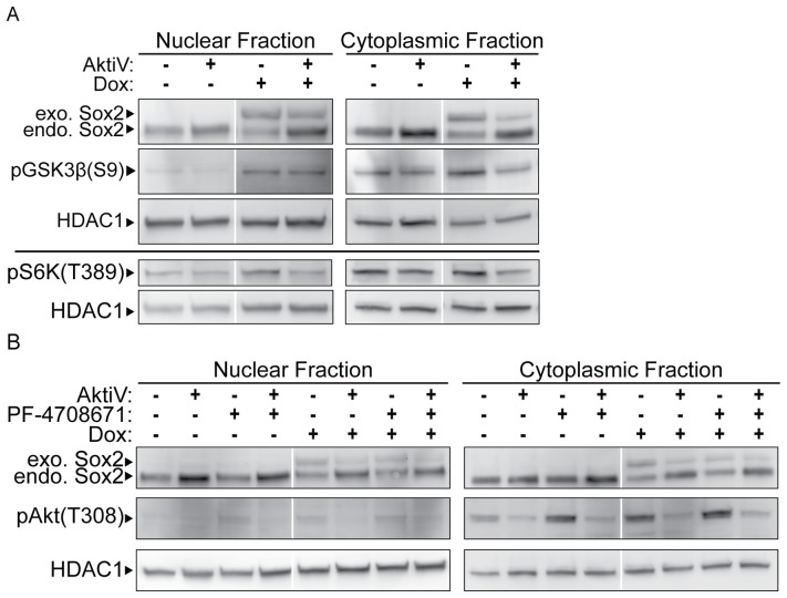
Figure 5. Effects of elevating Sox2 on phosphorylation of GSK3β and S6K.
i-OSKM-ESC were seeded at 1.5×10⁶ per 100 mm dish with or without 4 μg/ml Dox for 24 hours. (A) After the initial 24 hours, the cells were refed with fresh medium with or without 4 μg/ml Dox, and treated with 5 μM AKTiV for an additional 24 hours where indicated. 48 hours after the cells were plated, nuclear and cytoplasmic protein extracts were prepared and equal amounts of nuclear and cytoplasmic protein were loaded into each well of an SDS-PAGE. Western blot analysis was performed by sequentially probing for pGSK3β(S9), Sox2 and HDAC1 (top) or pS6K(T389) and HDAC1 (bottom). (B) After the initial 24 hours, the cells were refed with fresh medium with or without 4 μg/ml Dox, and treated with 5 μM AKTiV and/or PF-4708671 for an additional 24 hours where indicated. As in A, nuclear and cytoplasmic extracts were prepared from the cells and western blot analysis was performed by sequentially probing for pAKT(T308), Sox2 and HDAC1. In each case, HDAC1 served as a protein loading control.

