Figure 5.
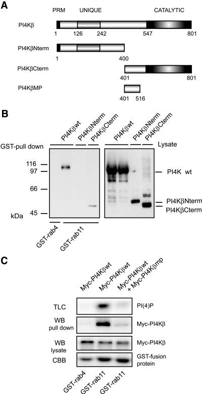
The rab11-binding site is located in the C terminus of PI4Kβ. (A) Schematic representation of PI4Kβ mutants. PRM, proline-rich motif; UNIQUE, lipid kinase unique domain; CATALYTIC, catalytic domain. (B) COS-1 cells were transfected with 5 μg cDNA encoding Myc-tagged PI4Kβwt (wild-type), PI4KβNterm (N-terminal domain, aa 1-400), or PI4KβCterm (C-terminal domain, aa 401-801). After 2 d, cells were lysed in 1 ml of lysis buffer, and GST pull-down assays were performed as described in the MATERIALS AND METHODS by using 5 μg of GST-rab4 or GST-rab11. Proteins bound to GSH beads were analyzed by Western blotting by using anti-Myc (GST pull down). Expression of the different constructs was analyzed by Western blotting by using anti-Myc (Lysate). (B) COS-1 cells were transfected with 5 μg of cDNA encoding Myc-tagged PI4Kβwt, or Myc-tagged PI4Kβwt in combination with Myc-tagged PI4Kβmp (middle piece, domain aa 401-516). GST pull-down assays were performed as in A. Proteins bound to GSH beads were split, either analyzed by Western blotting by using anti-Myc (WB, pull down) or subjected to an in vitro lipid kinase assay (TLC). As controls, PI4Kβ present in cell lysates was detected by Western blotting (WB, lysate), and the GST-fusion proteins were stained with Coomassie Brilliant Blue (CBB).

