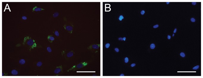
Figure 9. Cell staining with soluble M3-H12 Predator antibody.

The cell lines HVBP (A) and HMEC-1 (B) were stained with selected M3-H12 antibody and detected with Protein A conjugated with Alexa Fluor 488 (green). The cell nuclei were stained with DAPI (blue) (scale bar, 20 µm).
