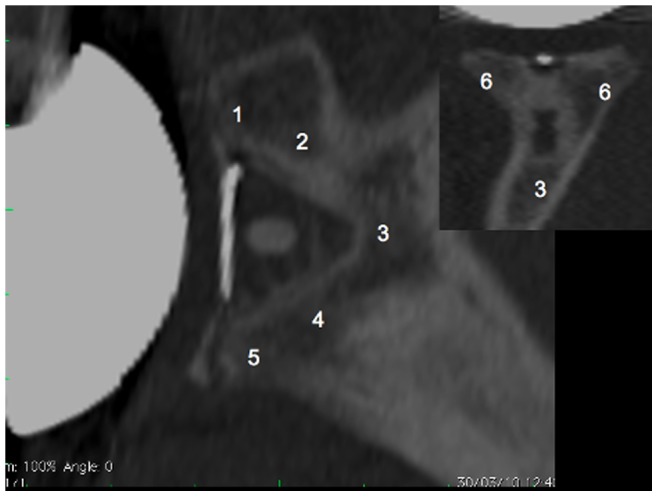
Figure 4. Sagittal and coronal views of the glenoid bone including the implant.

The numbers indicate the six zones used in the Mole Score to assess the level of radiolucent lines in the fixation of the glenoid component. Zone 1: fixation area of the superior part of the glenoid component base plate; Zone 2: fixation area of the superior part of the keel; Zone 3: fixation area of the tip of the keel; Zone 4: fixation area of the inferior part of the keel; Zone 5: fixation area of the inferior part of the glenoid component base plate; Zone 6: fixation area of the central part of the glenoid component base plate. Each zone is scored between 0 and 3 points according to the level of radiolucent lines observed and the Mole Score is the sum of these scores.
