
Figure 6. Morphological changes of human endothelial progenitor cells (EPCs) isolated from peripheral blood, and cultured for 7 days.
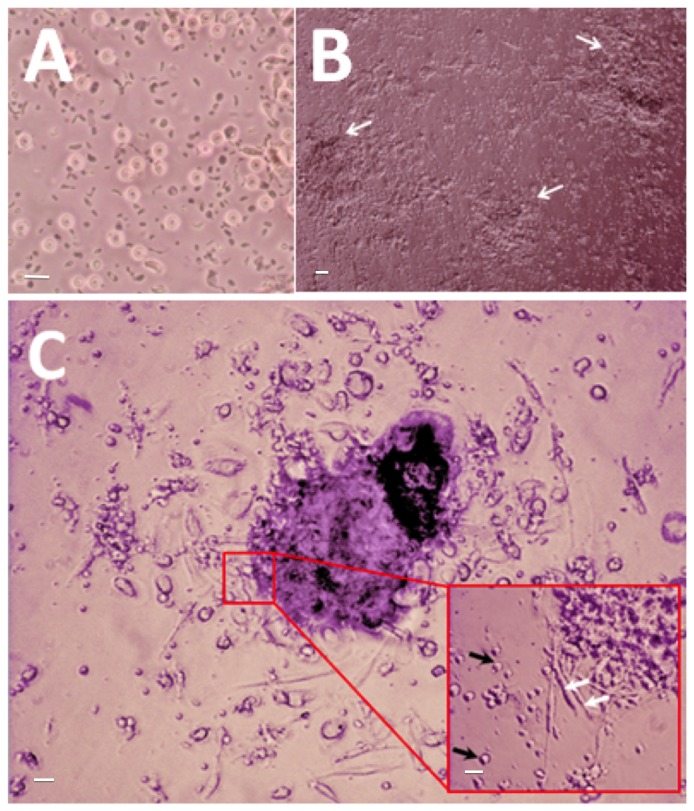
(A) Peripheral blood mononuclear cells (PBMCs) containing a heterogenous population of EPCs, monocytes, and granulophages, plated on normal uncoated tissue culture plate on day 1 (40x magnification). (B) White arrows point towards EPC colonies observed at day 7 (10x magnification). (C) An EPC colony at day 7, defined morphologically as central cluster of rounded cells surrounded by multiple spindle-shaped cells (20x magnification). Inset: Black arrows point to multipotent stem cells; white arrows point to EPCs (40x magnification). Scale bar represents 20 µm.