
Figure 12. Platelet activation assay.
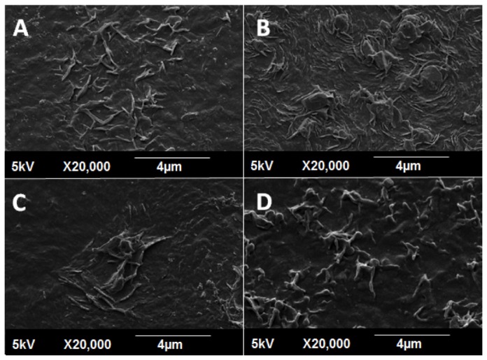
SEM images (2000x magnification) of adhered platelets show distinctly different morphological appearances. (A) Collagen, (B) POSS-PCU, (C) POSS-PCU-FS, (D) POSS-PCU-FS+CD34. Adhered platelets found on collagen-coated surfaces (positive control, A), showed the highest degree of activation, with formation of distinct pseudopodia and hyaloplasm spreading. Platelets adhering on POSS-PCU (B) and POSS-PCU-FS (C) were mostly dendritic-spread, with prominent pseudopodia as well as some flattening. Those adhered to POSS-PCU-FS+CD34 (D) remained dendritic with a clear spherical body and without any evident flattening.
POSS-PCU-FS: POSS-PCU with fumed silica anchors, POSS-PCU-FS+CD34: POSS-PCU biofunctionalized with anti-CD34 antibodies.