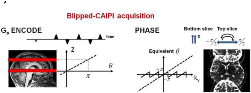
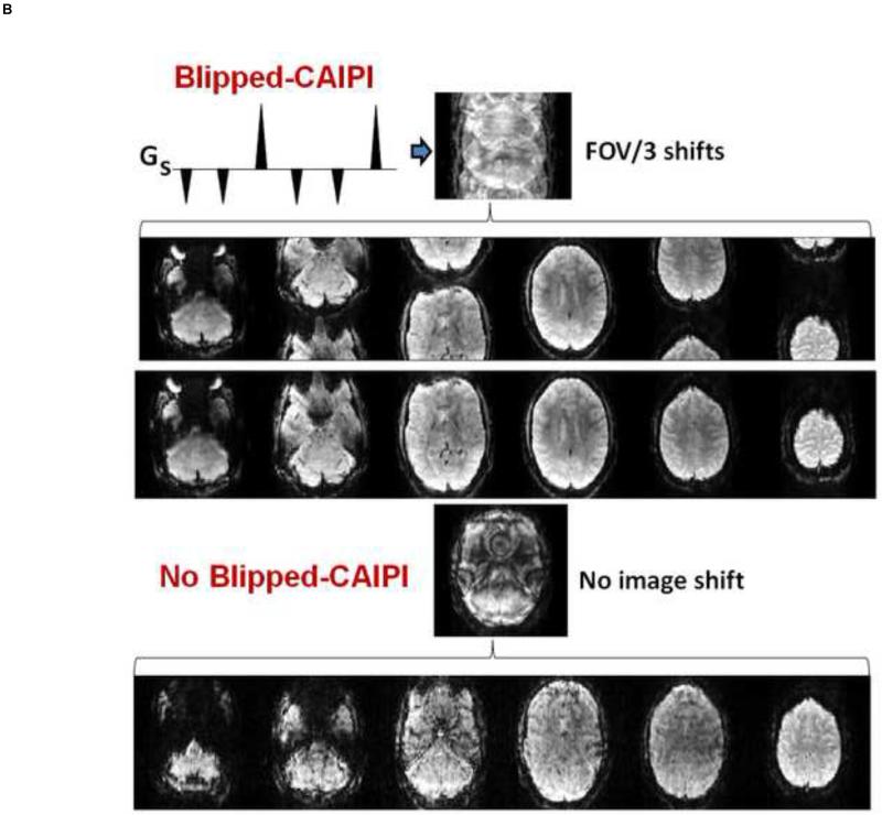
Figure 6.
A) provides details of the Blipped-CAIPI acquisition scheme. The Gs gradient pulses shown are played out during the echo train readout and modulate the phase of the echoes as a function of slice position on the slice axis. The images become shifted in the field of view (FOV/2) on the phase encoded axis. Adapted from [11]. B) Shown are FOV/3 inter-slice image shift in MB-6 simultaneous excited slices. The inter-slice shifted collapsed image is shown with its corresponding de-aliased images and final image process corrected images. Without blipped-CAPPI (bottom) there is superimposed alignment of the collapsed image data resulting in poorer image quality from image cross-talk and higher g-factor yielding lower SNR.
Modified from: K. Setsompop, J. Cohen-Adad, B.A. Gagoski, T. Raij, A. Yendiki, B. Keil, V.J. Wedeen, and L.L. Wald, Improving diffusion MRI using simultaneous multi-slice echo planar imaging. Neuroimage 63 (2012) 569-580.


