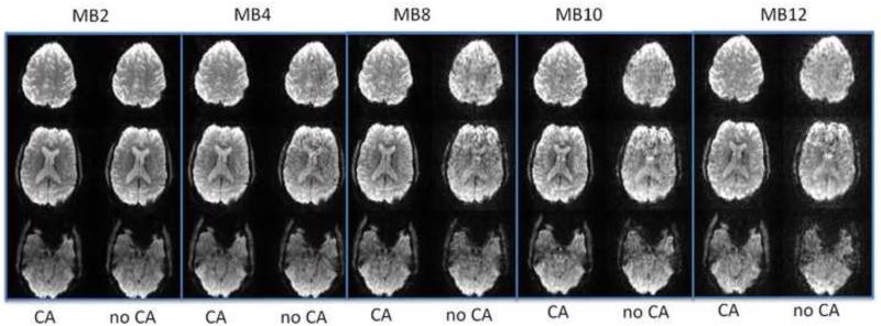
Figure 7.
Comparison of SMS EPI with blipped-CAIPI controlled aliasing (CA) and without (no CA). At low acceleration of MB2 images look about the same but at MB-4 and higher MB-8 there is severe artifact with no CA and higher g-factor in images. Each column shows 3 of 36-40 images acquired in separate scan of the brain with all parameters except MB-factor held constant: TR=2s, TE=36 ms, 70°, isotropic 2.5 mm voxels. The actual minimum achievable scan time of the brain could be reduced by shortening TR from the time required for normal EPI ~3s divided by MB down to ~250 ms at MB12. Shorter TR requires reducing flip angle to reduce SAR, which also changes image contrast. (provided by Liyong Chen and David Feinberg)

