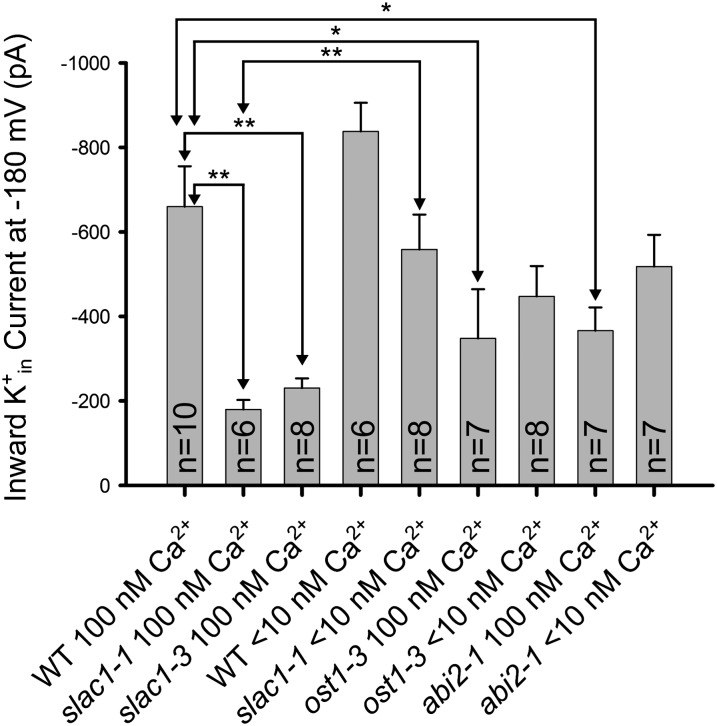
Figure 1.
K+in channel current activity is reduced in the stomatal closing impaired mutants slac1, ost1, and abi2-1 and this K+ channel down-regulation is rapidly reversed in slac1 guard cells by lowering [Ca2+]cyt to less than 10 nm. Average K+in channel current magnitudes at −180 mV are shown for wild-type Columbia-0 (WT) and the slac1, ost1, and abi2-1 alleles. The concentrations of buffered [Ca2+]cyt concentrations are indicated. Whole-cell patch clamp recordings were performed on guard cell protoplasts at the indicated cytosolic free Ca2+ concentrations. Note that an abi2-1 allele in the Columbia-0 accession was analyzed (Nishimura et al., 2004). Error bars (sem) for the indicated number of guard cells analyzed are depicted. K+in channel current magnitudes in slac1 recovered at less than 10 nm [Ca2+]cyt compared with 100 nm [Ca2+]cyt (P < 0.005). Statistical analyses showed significant down-regulation of K+in channel current magnitudes in the slac1-1, slac1-3 (P < 0.001), ost1 (P < 0.04), and abi2-1 (Columbia-0; P < 0.02) mutants compared with wild-type guard cells at 100 nm free [Ca2+] in the cytosol. Small but statistically nonsignificant differences for comparisons of K+in channel current magnitudes in response to lowering [Ca2+] from 100 nm to less than 10 nm for ost1-3 (P value = 0.525) and abi2-1 (P value = 0.109) were found. *P < 0.05; **P < 0.01. Unpaired Student’s t tests were applied to assess significance. Data from WT < 10 nm and slac1-1 < 10 nm are from Laanemets et al., 2013. Methods were as described in Laanemets et al., 2013 (see Supplemental Text S1).

