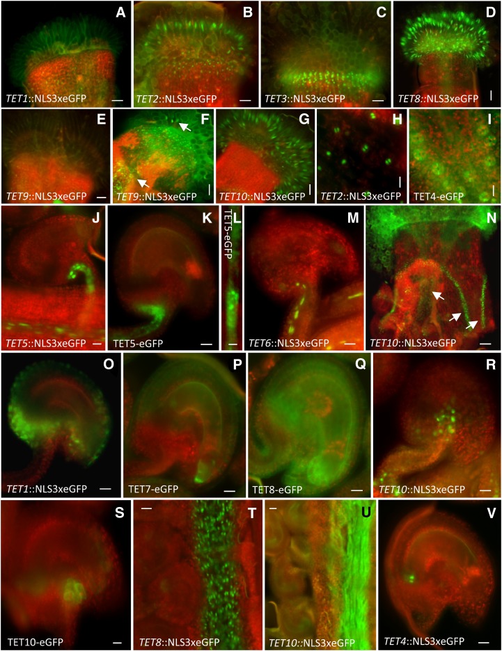
Figure 5.
Representative images of Arabidopsis tetraspanin expression patterns in diploid reproductive tissues. Transcriptional GFP fusions of various tetraspanins expressed in stigmatic carpel and papilla (a–g). TET9 expression in stigmatic papilla before (e) and after pollination (f). Arrows in (f) point to TET9 expression in transmitting tissue and papilla. Localization of TET2 transcriptional fusion (h) and expression of TET4 translational fusion (i) in carpel stomata. Localization of TET5 and TET6 in vascular tissues (j–m). Magnification of a vascular strand showing punctuated deposition (l). TET10 transcriptional fusion expressed in transmitting tissue (after pollination) and valve margins of the carpel (n). Transcriptional and translational fusions of various tetraspanins in ovule tissues after pollination (o–s). TET8 transcriptional fusion (t) and TET10 translational fusion (u) in the transmitting tract after pollination. Localization of transcriptional fusion of TET4 in ovules (v). Green fluorescent refers to GFP, and red fluorescent signal represents tissue autofluorescence. Bars = 50 μm (a–g, t, and u), 100 μm nN), 25 μm (j, k, m, o–s, and v), and 10 μm (h, i, and l).

