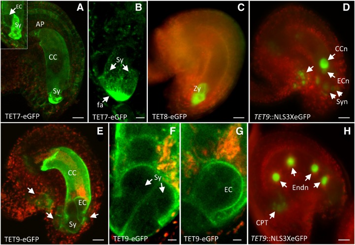
Figure 6.
Representative images of Arabidopsis tetraspanin expression patterns in female gametophyte and gametes. Expression of TET7 translational fusion in the plasma membrane of central cell (CC), antipodals (AP), and accumulation in the filiform apparatus (fa) of synergids (Sy). Inset shows an optical section pointing to egg cell lacking TET7 expression (a). Optical section of filiform apparatus showing TET7 granules distributing in a gradient within synergids (b). Representative image of TET8 translational fusion in atypical zygotes (Zy; c). TET9 expression before fertilization (d), showing egg cell nucleus (ECn), central cell nucleus (CCn), and synergid nuclei (Syn); expression of TET9 translational fusion in ovule and female gametophytic cells. Arrows point to individual cells in the micropyle and flanking the egg cell apparatus (e). Detail of TET9 translational fusion in the membrane of both synergids (f) and egg cell (EC; g). TET9 transcriptional fusion after fertilization (h) showing expression in endosperm nuclei (Endn) and chalazal proliferating tissue (CPT; arrows). Green fluorescent signal refers to GFP, and red fluorescent signal represents tissue autofluorescence. Bars = 20 μm (a and c–f), 10 μm (b), and 5 μm (g and h).

